

1. Advanced imaging technology ensures fast, precise scans with superior image quality.
2. Suitable for scanning equine, bovine, ovine, swine, feline, canine, and more.
3. Ideal for diagnosing the abdomen, obstetrics, cardiology, small parts, vascular, tendon, etc.
4. Comprehensive probe options meet diverse clinical needs.
5. Powerful measurement software offers extensive diagnostic support.
6. Smart, mobile design for easy portability.
7. Built-in batteries enable long-lasting outdoor use.
8. Efficient workflow ensures easy, comfortable operation.
- General Information
A brand-new ultrasound diagnostic platform with Innovations in areas of digital electronics achieves a new level of ultrasound diagnostic precision and higher diagnostic confidence.
A revolutionary workflow control is provided with the user-centric architecture of the new software platform.
- Main technical parameters and functions
2.1 Technical platform
★linux +ARM+FPGA
2.2 Channels and elements
Number of physical channels: ≥40
★Number of probe array element number:≥80
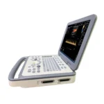
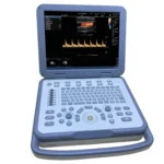
2.3 Size and weight
Host:
size:370mm (width) * 350mm(depth) * 70mm(thickness)
weight: 5.5kg(with no probe)
Including package:
Size: 455mm(width) ×455mm(depth) × 210mm(thickness)
weight: 9kg (host + 2 probes + power cable + adapter + manual + carton + foam)
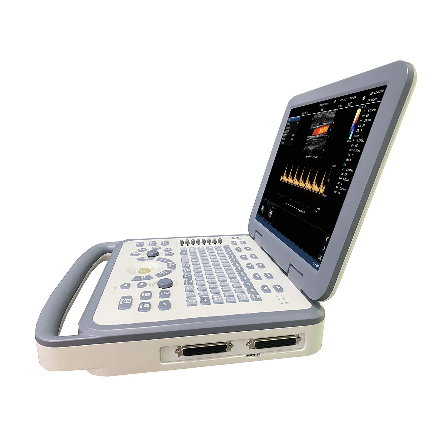
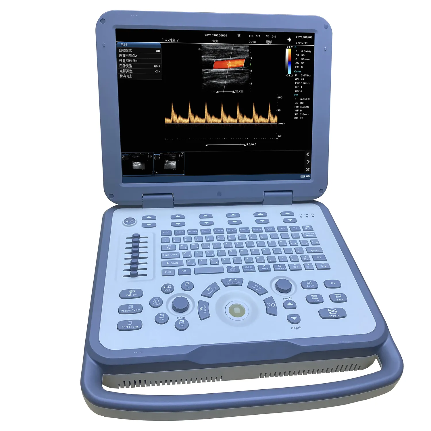
2.4 Monitor
15-inch, high resolution, progressive scan, Wide Angle of view
Resolution:1024*768 pixels
Image display area is 640*480 mm
2.5 Hard disk
★Internal 128GB hard disk for patient database management
Allow storage of patient studies that include images, clips, reports and measurements
2.6 Transducer Ports
Two active universal transducer ports that support standard(curved array, linear array)Probe, Unique industrial design provides easy access to all transducer ports
2.7 Probe available
3C6A: 3.5MHz/R60/80,Convex array probe
7L4A: 7.5MHz/L38mm/80. Convex array probe
6I7A: 6MHz/L64mm/80, Intrarectal Linear array probe
6C15A: 6.5MHz/R15/80, Micro convex array probe;
2.8 Imaging modes
B-mode: Fundamental and Tissue harmonic imaging
Color Flow Mapping (Color)
★B/BC Dual Real-Time
Power Doppler Imaging (PDI)
PW Doppler
M-mode
2.9 frequency number
B/M:Fundamental wave,≥3;harmonic wave: ≥2
Color/PDI:≥2
PW: ≥2
2.10 Cine
B mode: ≥5000 frames
B+Color/B+PDI mode: ≥2300 frames
M、PW: ≥ 190s
2.11, Image zoom
available on live, 2B, 4B and reviewed images
up to 10X zoom
2.12, Image save
format:
BMP, JPG, FRM(single image);
CIN, AVI(multiple images)
Support DICOM, conform to DICOM3.0 standard
★ Built-in workstation, supports patient data search and browse
2.13 Language
Support Chinese, English, Spanish, French, German, Czech, Russian languages.
Can be easily extended to support other languages
2.14 Battery
Built-in large-capacity lithium battery, working condition. Continuous working time ≥1 hour. The screen provides power display information
2.15 Other functions
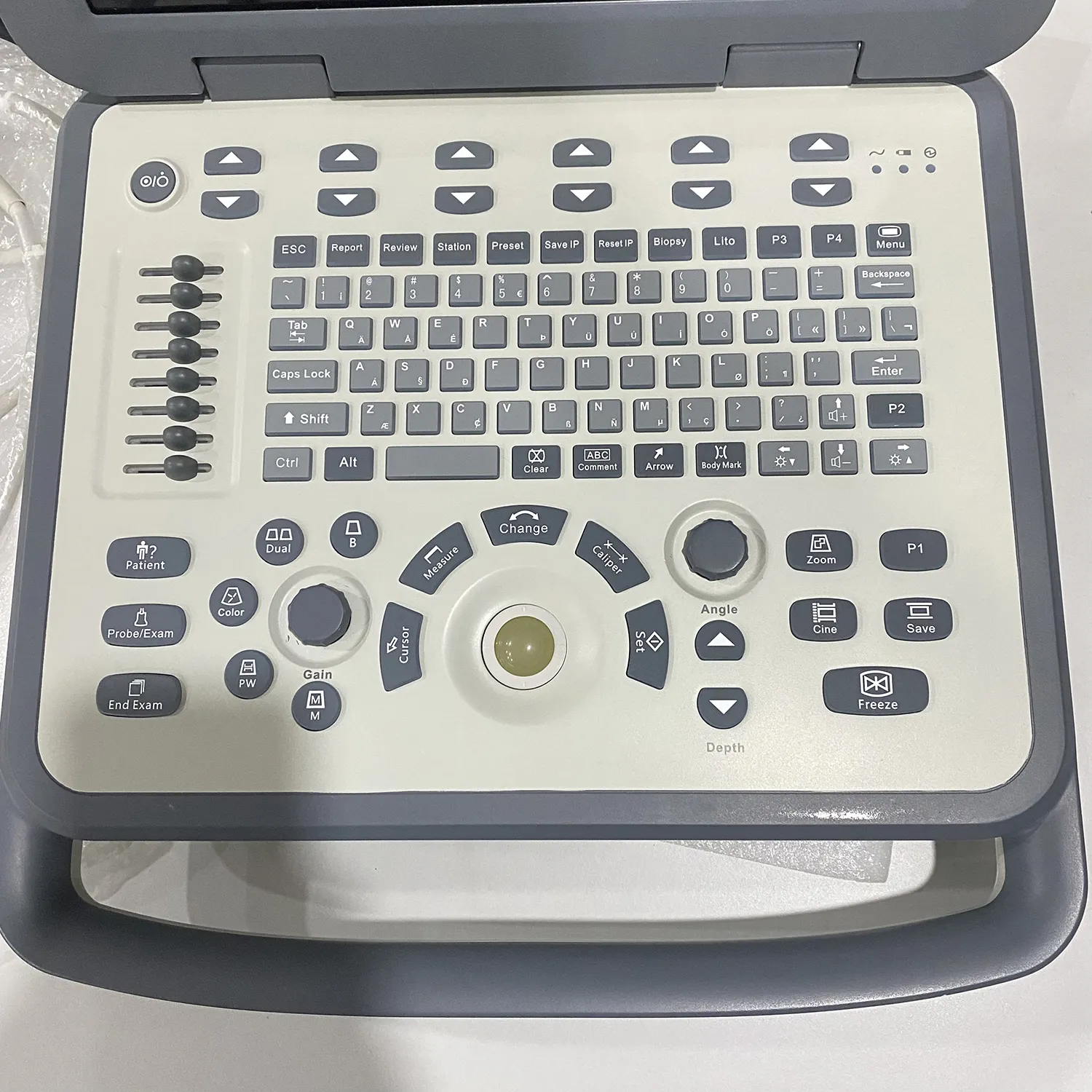
Comment, BodyMark, Biopsy, PICC, ★Lito, ★IMT, ★Report template, ★Support USB mouse, etc
Imaging Parameters
3.1 B mode
Up to four frequencies in fundamental imaging
Up to two frequencies in Tissue harmonic imaging (probe dependent)
Dynamic range | 0-100%, 5% step |
Speckle Reduction | 8 levels(0-7) |
Scan Density | H. M. L |
Gain | 0~100 %, 2% step |
TGC | eight TGC controls |
Frame Average | 8 levels(0-7) |
Line Average | 8 levels(0-7) |
Edge Enhance | 8 levels(0-7) |
Gray Maps | 15 types(0-14) |
Pseudo color Maps | 7 types(0-6) |
Thermal Index | TIC, TIS, TIB |
2B, 4B formats | / |
Invert (U/D) and transposed (L/R) | / |
Focus Number | 4 |
Focus Depth | 16 levels(depth and probe dependent) |
FOV | 5 levels |
Image depth up to 35 cm in 0.5~4cm increments (depth dependent) | |
Phase inversion harmonic imaging technique is available for all probes | |
3.2 Color mode
Frequency | 2 levels |
Gain | 0~100%, 2% steps |
Wall filter | 8 levels(0-7) |
Sensitivity | H, M, L |
Flow | H, M, L |
Packet Size1 | 5 levels(0-4) |
Frame Average | 8 levels(0-7) |
Post Proc | 4 levels(0-3) |
Invert | On/Off |
Baseline | 7 levels(0-6) |
Color Maps | 4 levels(0-3) |
Color/PDI Width | 10%-100%, 10% |
Color/PDI Height | 0.5-30cm(probe dependent) |
Color/PDI Center Depth | 1-16cm(probe dependent) |
Steer | ±12°, ±7°(linear probe) |
3.3 PDI mode
Frequency | 2 levels |
Gain | 0~100%, 2% steps |
Wall filter | 8 levels(0-7) |
Sensitivity | H, M, L |
Flow | H, M, L |
Packet Size1 | 5 levels(0-4) |
Frame Average | 8 levels(0-7) |
Post Proc | 4 levels(0-3) |
Invert | On/Off |
Baseline | 7 levels(0-6) |
PDI Maps | 2 levels(0-1) |
Color/PDI Width | 10%-100%, 10% |
Color/PDI Height | 0.5-30cm(probe dependent) |
Color/PDI Center Depth | 1-16cm(probe dependent) |
Steer | ±12°, ±7°(linear probe) |
3.4 PW mode
Frequency | 2 levels |
Sweep speed | 5 levels(0-4) |
Scale | 16 levels(0-15)(depth and probe dependent) |
Scale Unit | cm/s, KHz |
Smooth | 8 levels(0-7) |
Pseudocolor Maps | 7 types(0-6) |
Dynamic range | 24-100, 2 step |
Gain | 0-100%, 2% step |
Wall filter | 4 levels(0-3) |
Dynamic range | 24-100, 2 step |
Gain | 0-100%, 2% step |
Wall filter | 4 levels(0-3) |
Angle correction | -89°+89°, 1°step |
Gate size | 8 levels(0-7mm) |
Wall filter | 5 levels(0-4) |
Invert | On / Off |
Baseline | 7 levels |
Real-time auto Doppler trace: maximum velocity, mean velocity | |
3.5 M Mode
Frequency | Up to 3 fundamental and 2 harmonic imaging frequencies |
Edge enhance | 8 levels(0-7) |
Dynamic range | 0-100%, step 5% |
Gain | 0-100%, step 2 |
Gray Maps | 15 levels(0-14) |
Pseudocolor Maps | 7 (0-6) |
Sweep speed | 5 levels(0-4) |
3.6 image parameter save and restore
user can press one key to save image parameters on screen
Users can press one key to restore image parameters to default status.
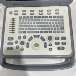
- Ergonomic Design
Frequently used controls center around the trackball
Control panel is backlighted, waterproof and antisepticised
Two USB ports are at the back of the system, which is more convenient for use
- Exam Modes
Abdomen
Obstetrics
Heart
Small parts
Vascular
Product Configuration
6.1 Standard Configuration
Host(Built-in 128G hard disk)
Adapter
6C15A Micro convex probe
7L4A linear array probe
User’s Manual
Power cable
6.2 Optional Accessories
3C6A convex array probe
6I7A Intrarectal Linear Array probe
USB report printer
B/W or color Video printer
Puncture rack
Trolley
Footswitch
- Suitable for scanning equine, bovine, ovine, swine, feline, canine, and more.
- Ideal for diagnosing the abdomen, obstetrics, cardiology, small parts, vascular, tendon, etc.
